|
There are several issues that affect our bulldog spines. We have
put together a collection of articles and web resources for information regarding this subject. To date there have been little
studies with our breed on this subject. Bulldogs are not often evaluated for their spines unless clinical signs are apparent.
Further focus is needed in this area for our breed. Until xrays are evaluated and submitted to researchers, little
data is collected on our very special bulldog spine.
Let's Talk Hemivertbrae
A couple of view points and information:
CLINICAL-RADIOLOGICAL STUDY OF THE VERTEBRAL
ABNORMALITIES IN THE ENGLISH BULLDOG
A.Volta, J.P. Morgan *, G. Gnudi, M. Bonazzi, M. Gazzola, S. Zanichelli , L. De Risio , G. Bertoni* Dpt. of Clinical
Sciences, School of Veterinary Medicine, Ross University, St. Kitts; Dpt. of Animal Health, Faculty of Veterinary Medicine,
University of Parma, Italy
Introduction : the aim of the study was to detect vertebral
abnormalities in a series of english bulldogs and to compare clinical and radiographic findings.
Materials and methods : a group of 38 english bulldogs underwent complete
physical and neurological examinations along with survey radiographs of the entire spine, prior to 4 months of age. From this
group, 23 dogs were re-evaluated both clinically and radiographically with:11dogs once, between 4 and 8 months, 4 dogs twice,
between 4 and 14 months, 5 dogs three times, between 4 and 27 months, 3 dogs five times, between 4 and 29 months.
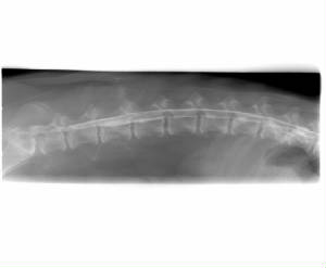
Results : radiographic investigation showed 148 thoracic hemivertebrae
in 37 dogs (97,3%). At least one segment was involved with the understanding that many dogs had multiple lesions. Five of
them (13,5%) showed mild neurological deficits, all consistent with a T3-l3 transverse myelopathy. The neurological signs
were noted earliest at 3 months in 3 dogs, 8 months in 1 dog, 27 months in 1 dog and were stable. Butterfly vertebra, as typically
described, was the most frequent type of anomaly (89affected vertebrae), followed by unilateral (30), dorsal (25) and ventral
(4) wedging of a vertebral body.
The most frequently affected vertebra was T9 (22 cases), followed by T7 (20), T10 (19), T8 (18), T12(16), T6
(15), T5 (11), T11 (10), T13 (9), T4 (6), T3 and T2 (1 each). It was difficult to obtain an exact number of segments and difficult
to determine if the lesions were primary or secondary. Other abnormalities included: thoracic spinous processes fusion in
4 dogs between t8 and T9 (3 cases) and between T7 and T8 (1) and thoracic and lumbo-sacral spondylosis (8 each). Coccygeal
anomalies (block and/or hemivertebrae) have been detected in all dogs.
Conclusion :
the study suggested that in this series of english bulldogs the occurrence of hemivertebra was very common, but rarely associated
with neurological deficits that required a treatment, at least during the first year of life.
|
|
What do they know we do not???
~*A great link and Article
about Hemivertebrae from the Boston Terrier Club.
~*A great link and Article
about Hemivertbrae from the French Bulldog Spine Data Base.
Typical Spine issues and the terminology
Congenital vertebral anomaly
Congenital vertebral anomalies are a collection of malformations of the spine in animals. Most are not clinically
significant, but they can cause compression of the spinal chord by deforming the vertebral canal or causing instability.
This condition occurs in the womb. Congenital vertebral anomalies include alterations of the shape and number of vertebrae.
Hemivertebrae
Among the congenital vertebral anomalies, hemivertebrae are the most likely to cause neurologic problems. They are wedge
shaped vertebrae, and therefore can cause an angle in the spine. The probable cause of hemivertebrae is a lack of blood supply
causing part of the vertebrae to not form. Hemivertebrae in dogs are most common in the tail, resulting in a screw shape,
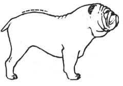
but can also occur in the thoracic vertebrae. Affected dog breeds include French and English Bulldogs, Pugs, and Boston Terriers. It is inherited in Yorkshire Terriers and German Shorthaired Pointers. The condition can cause death in very young Bulldog puppies.
Block vertebrae
Block vertebrae occur when there is improper segmentation of the vertebrae, leading to parts of or the entire vertebrae
being fused. It can lead to an angle in the spine, but there are usually no symptoms. The sacrum is a normal block vertebrae.
Butterfly vertebrae
Butterfly vertebrae have a cleft through the body of the vertebrae and a funnel shape at the ends. This gives the appearance
of a butterfly on an x-ray. It is caused by persistence of the notochord (which usually only remains as the center of the intervertebral disk) during vertebrae formation. There are usually no symptoms. Butterfly vertebrae occur most often in Bulldogs, Pugs, and Boston
Terriers.[1]
Transitional vertebrae
Transitional vertebrae have the characteristics of two types of vertebrae. The condition usually involves the vertebral
arch or transverse processes. It occurs at the cervicothoracic, thoracolumbar, or lumbosacral junction. For instance, the transverse process of the last
cervical vertebrae may resemble a rib. A transitional vertebrae at the lumbosacral junction can cause arthritis, disk changes, or spinal cord compression.
References
- ^ a Ettinger,
Stephen J.;Feldman, Edward C. (1995). Textbook of Veterinary Internal Medicine, 4th ed., W.B. Saunders Company.
|
 |
|
|
|
|
|
|
|
|
|